


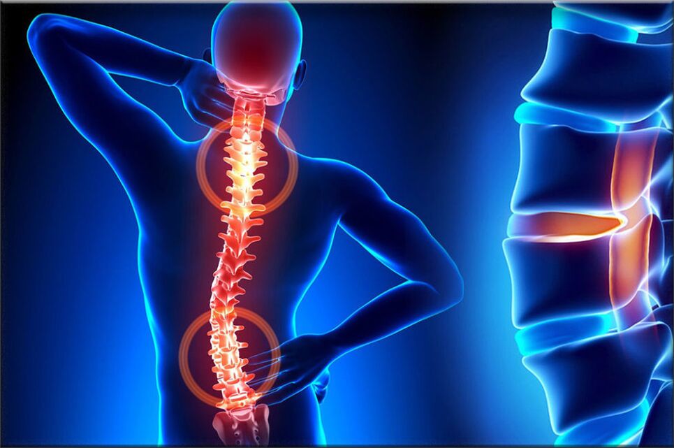
Osteochondrosis is one of the most common diseases of the musculoskeletal system, which manifests itself as a result of certain dystrophic changes in the vertebral cartilage, during this pathological process, the spinal disc is often affected. Its structure, which is an intervertebral cartilage disc, provides flexibility, and also allows the human spine to move, that is, it provides movement.
With osteochondrosis, a number of processes occur that cause degeneration of the vertebral discs, as a result of which they begin to lose elasticity and reduce the level of flexibility, and during this time the disc itself becomes quite flat. The distance between the two discs is reduced, while compressing the nerve endings and blood vessels and causing severe pain. The site of nerve compression begins to swell, resulting in increased pain and greater violation.
During the development of osteochondrosis, muscle structures and most organs of the body are often involved in this pathological process. This is due to the fact that during the maximum violation of the neurovascular bundle, blood circulation and movement of muscles and organs is disrupted. For example, the most common osteochondrosis is cervical osteochondrosis, which is accompanied by pain in the back of the head, nausea, dizziness, impaired vision and often tinnitus. The disease has become "younger": a century ago, osteochondrosis was a disease of people of gerontological age, and today young people are also susceptible to it.

The most vulnerable categories of people are those with disorders of metabolism and hormone levels of the body, as well as people with disorders of vascular-venous nature. This is due to the fact that the disease causes disruption of disc oxygenation. If qualified, timely steps are not taken to heal, then the edges of the affected intervertebral disc, which are compacted, will anatomically protrude beyond the boundaries of the spinal space, thus destroying the neurovascular bundles.
Therefore, patients are at risk of developing a herniated disc. The main cause of osteochondrosis is the uneven distribution of load on the spine, which leads to the fact that the structure of cartilage changes at points with excessive pressure. The nature of the disease depends on the degree and degree of damage to the affected disc. The intervertebral disc changes with age, as does our hair. A large injury or fracture of the spine can affect how it works. Casual clothing and certain types of vibrations can also accelerate the rate of spinal degeneration. In addition, evidence suggests that smoking increases the rate of spinal degeneration. Scientists have also discovered links between family members, highlighting the role of genetics in how quickly changes occur.
The disease can also be triggered by various factors:
- injuries, bruises;
- spinal muscular dystrophy;
- tension and curvature of the spine;
- weight lifting;
- prolonged stay in one position;
- metabolic diseases;
- lack of trace elements and vitamins - manganese, magnesium, zinc and vitamins D and F;
- hereditary tendencies;
- physical load;
- inactive lifestyle;
- radiation background;
- cold sores;
- congenital dystrophy;
- asymmetrical work of the spinal space muscles;
- stress, depression.
The cause of this osteochondrosis is only the assumption of scientists, the direct factors that cause the disease, science has not yet found, and we only discuss risk factors.
The first periodexpansion - characterized by early proliferation of the intradiscal nucleus pulposus (nucleus pulposus of the eccentric intervertebral disc, located next to the dorsal part of the vertebra).
The second periodcharacterized by the appearance of spinal segment instability. The pathological substrate is represented by a fibrous core of the disc affected by the degenerative process of retrieval and rupture of the posterior longitudinal ligament, pathological movement between the vertebrae develops.
The third periodthe development of the disease - total damage to the intervertebral disc, with the appearance of a "herniated disc" - dislocation and removal of fragments of the nucleus pulposus outside the intervertebral space.
If the disease has reached the third phase, then the process of destruction is already irreversible and can lead to profound disability.
Types of osteochondrosis
The evolution of osteochondrosis is slow, with exacerbations caused by spinal cord injuries, exercise, heavy loads, etc. The clinic depends on the location of the lesion.
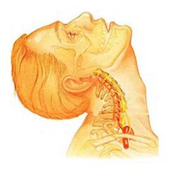
Osteochondrosis of the cervical spinehas local and isolated symptoms of advanced form - with a strong root dominance, that is, it contributes to the development of severe radicular pain. Symptoms of osteochondrosis of the cervical spine are accompanied by varying degrees of dysfunction, sometimes manifested in limitation of movement of the cervical spine and sudden block of function. Headaches can be exciting and paroxysmal naturally with irradiation to the interscapular area or shoulder area. In the acute period, the patient is diagnosed with an attack of pain in the neck, which prevents and restrains the movement of the head and neck. In addition to severe discomfort, pain syndrome can be accompanied by dizziness, insomnia, pain, loss of appetite, depression, eye and pharyngeal diseases.
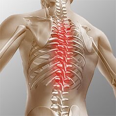
Osteochondrosis of the chest. . . Clinical manifestations are due to localized lesions and the process of destruction of nerve root structures. Thoracic osteochondrosis has a significant pain syndrome, which can have chronic or acute back pain with chest discomfort and limited muscle contraction, to right-muscle atrophy. Chest pain can manifest itself as diffuse, intercostal, and neuralgic. Palpation increases the axial rotation of the vertebral body. The disorder corresponds to the degree of root irritation from Thl1 to Thl2, and can manifest itself as angina pectoris, which is reflected in dysfunction of the function of the liver and gastrointestinal tract. Disorders of the genitourinary system and genital area often occur. Patients noted sensory disturbances such as paresthesia, superficial and profound sensitivity was significantly reduced.
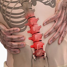
Lumbar osteochondrosis. . . It is characterized by abdominal reflexes and lower leg dysfunction. During the development of neurological disorders, muscle weakness in the legs and dysfunction of the pelvic organs can occur. Osteochondrosis is characterized by assessing damage to the sitting process. The more advanced the stage of development of the lumbar vertebral lesion, the shorter the length of time the patient can sit. The lumbar form is characterized by chronic and acute back pain, paravertebral muscle spasm, and secondary myofascial syndrome. The pain radiates to the back and posterior ilium.
Depending on the localization of the pathological process of osteochondrosis, the disease can cause the patient to experience a violation of superficial sensitivity (tactile, thermal). Also characteristic are reflex changes (e. g. , Achilles reflex absent), muscle wasting, muscle tone disorders, autonomic disturbances (pallor, redness of the skin, trophic changes in the nails, skin hypothermia at the distal end), sphincter dysfunction and sexual dysfunction.
Clinical picture
Diagnosticsstarting with a history and a complete physical examination. The doctor asks questions about the symptoms, how the disease interferes with the daily activities of the patient. Also, specialists are interested in identifying positions and activities that emphasize or reduce pain levels.
The doctor then examines the patient, checking the position and distance of movement in the spine, thus determining the movement that is causing the pain. Skin sensitivity, muscle strength and reflexes were tested equally. Based on medical history and physical examination, the doctor determines which technique will help.
Radiographs rarely aid diagnosis, no more than 30% of radiographic images show abnormalities in the early stages of disease development.
However, if the symptoms are severe and the disease is already in the second or third stage, a defect in one or more intervertebral discs can be seen in the picture. They can be penetrated by osteophytes between the vertebrae and joints.
If additional information is required, magnetic resonance imaging is prescribed. MRI is used to look at the soft tissues of the body. This is useful if the tissue core absorbs water, or if there are cracks in the disc. MRI can show problems in other soft tissues, such as the spinal cord.
Discography can help in diagnosis. This examination is performed using contrast agents, each of which is injected into one or more discs. Subsequent projections on the radiography provide useful information about the condition of the disc.
Treatment of osteochondrosis, depending on its type
Non -surgical treatment of osteochondrosis
If possible, doctors prefer non -surgical treatment. The most important thing in non -surgical treatment is to relieve pain and other discomforts so that the patient can continue a comfortable standard of living as much as possible.
Doctors rarely prescribe bed rest for patients with osteochondrosis. Patients are encouraged to live in natural movements when pain is not a concern. If the symptoms are severe, a few days of sleep rest may be prescribed.
When the spine is transplanted, an elastic belt is sometimes prescribed, which is worn no more than 2-4 days to prevent atrophy of the back muscles.
Osteopathy sessions provide serious relief to osteochondrosis.Doctor of osteopathynot only diagnoses problem areas, but also relieves pain in 1-2 doses, relieves the general condition of the body and "tightens" the visceral organs.
Patients can be given medication to control symptoms and resume normal activities over a long period of time. If symptoms continue to restrict the patient’s activity, a conventional physician may recommend an epidural steroid injection.
Steroids are powerful anti-inflammatory, helping to relieve pain and inflammation. Nonsteroidal anti-inflammatory injections are injected into the space around the spinal roots. This site is called the epidural space. Some doctors inject steroids only. However, it is most often combined with other drugs. Basically, steroids are prescribed only when other medications are ineffective, but osteopathy almost always helps.
In addition, patients often work with a physical therapist. After assessing the patient’s condition, the therapist prescribes exercises to reduce symptoms. The exercise program aims to increase flexibility and is useful for training the abdominal and back muscles to allow for movement with the least amount of pain.
Surgery
People with osteochondrosis usually do not need surgical treatment. In fact, only 1-3% can be controlled. The surgeon prescribes a non-surgical treatment, i. e. craniosacral osteopathy, as rehabilitation therapy, at least 3 months before considering surgery. If after 3 months of non -surgical treatment there are no results, there is only a reason that indicates a surgical procedure.
Basic surgical procedures
Dysectomy
This procedure aims to remove part or all of the disc in the lumbar region. Surgeons usually perform surgery through incisions in the lumbar region. Before removing a herniated disc, it is necessary to remove several plates.
Today, surgery has mastered minimally invasive techniques that require only small incisions in the lumbar region. Proponents of this method claim that it is safe. They also believe that the procedure can prevent scarring around nerves and joints and help patients recover faster.
Combined
This is an intervention that combines two or more bones into one, preventing the ends of the bones and joints from wearing and tearing.
Recovery
Doctors may recommend that patients see a physical therapist several times a week for 4-6 weeks. In some cases, patients need extra help.
The first year of treatment is needed to control symptoms. The therapist will work with you to find positions and movements that relieve pain. Hot, cold, ultrasound, and electrical stimulation can be prescribed to relieve muscle pain and cramps. Sequences or forms of soft tissue mobilization can also be used. This procedure helps the patient perform movements easily.
Typically, tailoring treatment helps restore the sensitivity of the nerves and muscles of the spine, reduce pain and increase mobility.
The main goal of therapy is to teach patients how to perform manipulations to avoid problems in the future. Patients will be given some exercises to increase flexibility. Patients will also be given strategies to help if there are recurrent symptoms.
Everyone should study and consider all types of osteochondrosis to prevent the development of this disease in himself and loved ones. However, treatment of a crushed vertebra is not possible, the therapy aims to relieve pain symptoms and achieve long -term remission. You also need to remember simple but effective rules:The best antidote is prevention. . .
Prevention of osteochondrosis
Prevention is simple enough - it’s a healthy diet, regular muscle activity, daily morning warm -ups, a healthy and active lifestyle and monthly visits.osteopathic sessionsfor the correction and elimination of musculoskeletal tension. Adhering to these rules is enough to never face such problems and avoid adverse symptoms and lifelong treatment.