


Cervical osteochondrosis is a disease in which the vertebral and intervertebral discs are affected. Cervical osteochondrosis refers to a deformed dorsopathy. Invocular changes in the disc were observed at the age of 20 years. At the same time, they become more sensitive to loads, less elastic, and lose lubricating fluid.
Often, pathology occurs in the elderly, but currently there is a significant increase in incidence among children and adolescents. Neurologists identify cervical osteochondrosis using the latest diagnostic studies. After clarifying the diagnosis, complex therapy is carried out with the most effective medications, physiotherapy procedures and innovative methods of physical rehabilitation.
The name of the disease consists of two Greek terms "osteon" (bone) and "chondros" (cartilage). Cervical osteochondrosis begins with changes in the center of the disc. The intervertebral disc loses moisture, decreases in size, this leads to the concentration of the vertebral body and the violation of nerve roots with ducts. The vertebrae receive nutrients from the surrounding tissues, which are harmful to the body. Compression of nerves and blood vessels leads to spasm of the protective muscles, which, as the disease progresses, becomes a cause of pain.

Which doctor treats this disease
Treatment of osteochondrosis is the field of activity of neurologists. However, when symptoms of neck osteochondrosis appear, it is possible to consult a general practitioner. A neurologist will choose the medication for cervical osteochondrosis that has the least pressure on the body, which is important for drug therapy.
To determine the presence of pathological processes in cartilage tissue and cervical osteochondrosis, the patient is referred for a comprehensive examination. Tactics on how to treat cervical osteochondrosis are being developed in accordance with the results of the investigation.
Interdisciplinary collaboration also enables the treatment of patient -owned comorbidities. In addition, patients receive full informational support: treatment plans, citations on the cost of services, provision of information on specialist consultations and diagnostic measures.
Cause
Cervical osteochondrosis develops under the influence of various provoking factors. No definite cause of cervical osteochondrosis has been identified. The disease is often associated with metabolic disorders and vertebral aging.
Researchers suggest that cervical osteochondrosis develops for the following reasons:
- Excessive pressure on the spine. High load on the spine is observed when wearing the wrong shoes, flat feet, obesity, prolonged sitting position;
- Metabolic disorders. Lack of vitamins, minerals, disorders of calcium metabolism can be the cause of degenerative processes in the vertebrae;
- Congenital and spinal anomalies and ligament apparatus (ligament thickening, lumbariization, sacralization);
- Pathology of the gastrointestinal tract, resulting in insufficient absorption of nutrients;
- Infection, intoxication;
- Injuries, bruises, fractures of the spine, as a result of which the blood supply and maintenance of the spinal canal are disrupted, resulting in disorders of their dystrophy;
- Pressure;
- Wear shoes with heels;
- Pregnancy, especially multiple pregnancies;
- Autoimmune lesions of connective tissue, abnormal collagen structure types 1 and 2;
- Occupational hazards (lifting heavy loads, prolonged vibration, working in a sitting position with constant head tilt);
- Atherosclerotic and other changes in the vertebral arteries;
- Curvature of the spine (kyphosis, scoliosis, kyphoscoliosis).
An important risk factor for the development of cervical osteochondrosis is heredity. This fact proves the presence of osteochondrosis in children, when the spine is not yet too much.
Degrees
Due to the special structure of the spine, it is able to perform its function. The main structural unit is considered to be the spinal motor segment (VMS). It consists of two adjacent vertebrae, the intervertebral disc and the musculoskeletal-ligament apparatus. Osteochondrosis leads to dystrophic degenerative processes, first in the intervertebral discs, then in the vertebrae. With the defeat of one vertebra, the performance of its function is provided by the nearby. This leads to increased load and loss of mobility of the affected segment.
In the development of cervical osteochondrosis, doctors distinguish several stages:
- The first stage of cervical osteochondrosis. Because the intervertebral disc lacks its own blood supply and receives nutrients from the surrounding tissues, the disc undergoes degenerative changes. Osteochondrosis in developmental stage 1 is characterized by destruction of the nucleus pulposus and rupture of the annulus fibrosus. Clinically, this is indicated by acute or persistent local pain in the neck (cervicalgia) and stiffness;
- Second degree osteochondrosis of the cervical spine. At this stage, destruction of the annulus fibrosus persists, pathological movements and vertebral instability appear. Patients complain of neck pain, exacerbated by physical exercise, tilting the head or in a certain position;
- The third stage of the disease is characterized by complete destruction of the fibrous annulus. The gelatin nucleus is not fixed. Herniated discs can occur and cause severe pain. At this stage, due to poor SMS fixation, curvature of the spine may form;
- In the fourth stage of the disease, the intervertebral disc is replaced by connective tissue, other adjacent segments are affected. Spondyloarthrosis, arachnoiditis develops. The joints become completely immobile - ankylosis develops. Bone tissue grows around the affected area - osteons are formed. With the fourth stage of cervical osteochondrosis, obvious symptoms are observed: severe pain radiating to the arms, sternum, to the area between the shoulder blades, disturbances of sensitivity.

Symptoms and Signs
Signs of cervical osteochondrosis in the early stages can be nonspecific: dizziness, headache, weakness, cracking during head movements. As the disease progresses, the following symptoms develop:
- Severe pain in the neck and shoulders;
- Numb hands;
- Dizziness;
- Blood pressure rises;
- Impaired movement coordination;
- Increased sweating.
There are several syndromes that appear with the development of pathological conditions of the muscles of the back and cervical spine:
- Cervical migraine syndrome.
- Vertebral artery syndrome.
- Hypertension syndrome.
- Heart syndrome.
- Radicular syndrome.
They occur when nerve endings are injured, arteries and veins are squeezed during the development of the disease. The most dangerous complication is considered to be vertebral artery syndrome. There is a violation of blood flow through the arteries that feed the brain and spinal cord. Patient hearing decreased, vision decreased, dizziness persisted. Patients may lose consciousness while driving due to a sharp violation of blood flow.
As a result of nerve compression responsible for the maintenance of the chest and diaphragm muscles, pain in the heart area appears, which is not associated with heart disease, but at the same time, tachycardia, arrhythmias and hypotension may develop. Vein compression leads to the development of hypertensive CSF syndrome. Increased intracranial pressure, nausea, vomiting, and severe headaches appear due to the disrupted outflow of blood from the brain.
As a result of squeezing the neck, radicular syndrome develops - severe pain appears in the neck, shoulders, shoulder blades, and back of the head. With this syndrome, the arm and neck area is numb. With cervical migraine syndrome, patients worry about severe pain in the occiput, which is often accompanied by nausea and vomiting.
Reflex syndrome occurs when the roots of the spine have not been affected. The patient complains of pain in the neck, head (especially the back of the head), in the arms on one or both sides. Reflex pain, unlike radicular pain, is not combined with sensory disturbances. Cervicalgia can be dull, painful. The sharp pain of the "lumbago" is called the cervicago. There are muscle cramps and pain, pain at the paravertebral point. Signs of cervical osteochondrosis increase in uncomfortable positions, with headaches, coughing, and physical exercise. Signs of epicondylosis, humeroscapular periarthrosis and shoulder-to-shoulder syndrome appear due to nerve impulses from the annulus fibrosus of the affected segment, which cause compensatory muscle spasm.
Radicular syndrome is accompanied by disturbances of motor activity and sensitivity. At the same time, nerves, blood vessels are disrupted, venous and lymph outflow at the pathological focus is disrupted due to the decline of the intervertebral canal. The pain in radicular syndrome is acute, intense. A common cause of spinal cord entrapment is the formation of a hernia. In the pathological focus area, muscle tone decreases. With radiculoischemia, in addition to nerves, the ducts are compressed.
If the phrenic nerve is involved in a pathological process, cardiac syndrome occurs. It manifests itself as a burning sensation, acute pain in the left side of the chest with irradiation to the arm, interduloid area. The name of this syndrome is due to the fact that the nature of the pain is similar to an attack of angina pectoris. The main difference between the pain in angina pectoris is that it is relieved after taking nitroglycerin, it can occur at rest and is combined with disturbances in heart rhythm (tachycardia, arrhythmias).
The signs of cervical osteochondrosis depend on the localization of the pathological process. With damage to the upper cervical vertebrae, the blood supply to the brain is disrupted due to compression of the cerebral arteries. This causes headaches (especially in the occipital area), dizziness, fainting, high blood pressure. Dizziness with cervical osteochondrosis is caused by decreased blood flow to the inner ear. Patients are also concerned about nausea, vestibular and ocular symptoms.
With joint lesions of the vertebrae, they talk about cervical osteochondrosis. The disease is indicated by the following symptoms:
- Dizziness;
- Pain in neck and arms;
- Tingling, a creeping sensation in the upper limbs;
- Intercostal neuralgia.
Diagnostics
Cervical osteochondrosis is a chronic disease that can lead to the formation of hernias and compression of the spinal cord. Therefore, it is important to establish a timely diagnosis and begin therapy. To identify cervical osteochondrosis, the following types of instrumental diagnostics are used:
- Spondylogography or X-ray of the spine. This research method is painless, very informative and does not require special training. X-rays of the spine allow you to assess its anatomical features and functions. In the picture, attention is paid to the structure of the vertebrae, their relationship to each other, the distance between them, the lumen of the spinal canal;
- Computed tomography - provides information primarily on the condition of bone tissue, allows you to identify narrowing of the spinal canal and herniated discs;
- Magnetic resonance imaging - allows you to determine changes in soft tissues. MRI images clearly show changes in the intervertebral disc and spinal cord.

Drug treatment
Treatment of osteochondrosis of the cervical spine consists of drug and non -drug therapy. Even after complete healing, neurologists take preventative measures to exclude recurrence of the disease. In the acute period, for the treatment of osteochondrosis of the cervical spine, the doctor prescribes drugs of the following pharmacological groups to patients:
- Analgesics are not narcotics. They are taken orally or injected intramuscularly to quickly achieve their effects;
- Non-steroidal anti-inflammatory drugs;
- Vitamin B in large doses.
Diuretics are used to reduce fluid retention in the roots of the spine and surrounding tissues. Antihistamines strengthen the analgesic action. Muscle spasms are relieved by muscle relaxants. With prolonged severe pain syndrome, neurologists perform nerve blockade.
To enhance metabolic processes in the intervertebral disc, chondroprotectors are used. The drug increases the content of glycosaminoglycans, increases the firmness, elasticity and shock absorption of the intervertebral disc.
Dizziness pills
Patients often experience dizziness with cervical osteochondrosis. To reduce it, doctors prescribe non-steroidal anti-inflammatory drugs. NSAIDs belonging to different groups differ in their mechanism of action and effects, therefore, only a qualified specialist can determine the appropriate drug.
It is important to remember that medications for osteochondrosis of the cervical spine should not be taken without a doctor’s appointment. Non-steroidal anti-inflammatory drugs have side effects, therefore, before prescribing them, the neurologist determines the presence of contraindications in the patient and the required dose. Medications for dizziness in cervical osteochondrosis can improve the quality of life of patients.
Injections for osteochondrosis
Injections for osteochondrosis of the cervical spine help relieve pain during exacerbations. With this method of drug administration, the effects occur quickly. Neurologists use a variety of injections.
The nurse injects the drug solution subcutaneously, intramuscularly, or intravenously. During the period of disease exacerbation, drugs given by injection, with cervical osteochondrosis, have an exclusively symptomatic effect.
Headache treatment
Headaches are symptoms that occur with a variety of disorders. However, cervical osteochondrosis is characterized by intense headache attacks. Head movements increase symptoms, therefore, to get rid of them, doctors prescribe analgesic tablets and non-steroidal anti-inflammatory drugs.
Non -drug therapy methods
Complex non -drug therapies of cervical spinal osteochondrosis include:
- Protective mode - when the root is pinched, the patient lies on a hard surface,
- Sort;
- Physiotherapy training;
- Spinal traction;
- Physiotherapy procedures.
Massage for cervical osteochondrosis is used to reduce pain and swelling, improve peripheral blood supply, and relieve muscle spasms. A contraindication to performing this procedure is the presence of acute pain. Massage the neck and back in the direction of lymph outflow. Particular attention is given to the interscapular and paravertebral zones.
Therapeutic gymnastics for osteochondrosis of the cervical spine aims to relieve muscle spasms and strengthen the muscular skeleton. Because vertebral instability often occurs in the cervical spine, the exercise therapy instructor conducts individual lessons, in which he or she teaches the patient to perform the exercises safely. A number of authors recommend holding physical therapy classes on Shants collars.
To improve the mobility of the cervical vertebrae, rehabilitation therapists recommend doing the following exercises:
- Bending and lengthening of the neck. Tilt your head forward toward your sternum, without pulling the shoulders forward, then backward. Hold sideways for 3 seconds, repeat each exercise 8-10 times;
- Neck dizziness. Turn your neck first to the left until it stops, then to the right, without changing the position of the shoulders and chin level;
- Lower your head until it stops. Then tilt your head back without changing your shoulder level. Hold the position for 5 seconds.
The following exercises have been developed to strengthen the neck muscles:
- Place your hands on the back of your head. Tilt your head back, leaning on your hands;
- Place your hands on the temporal area. While tilting the head, hold with your hands;
- Place your hands on your forehead, hold, tilt your head forward;
- Tilt your head to the side with your right hand, with your left hand behind your back. Repeat the exercise instead.
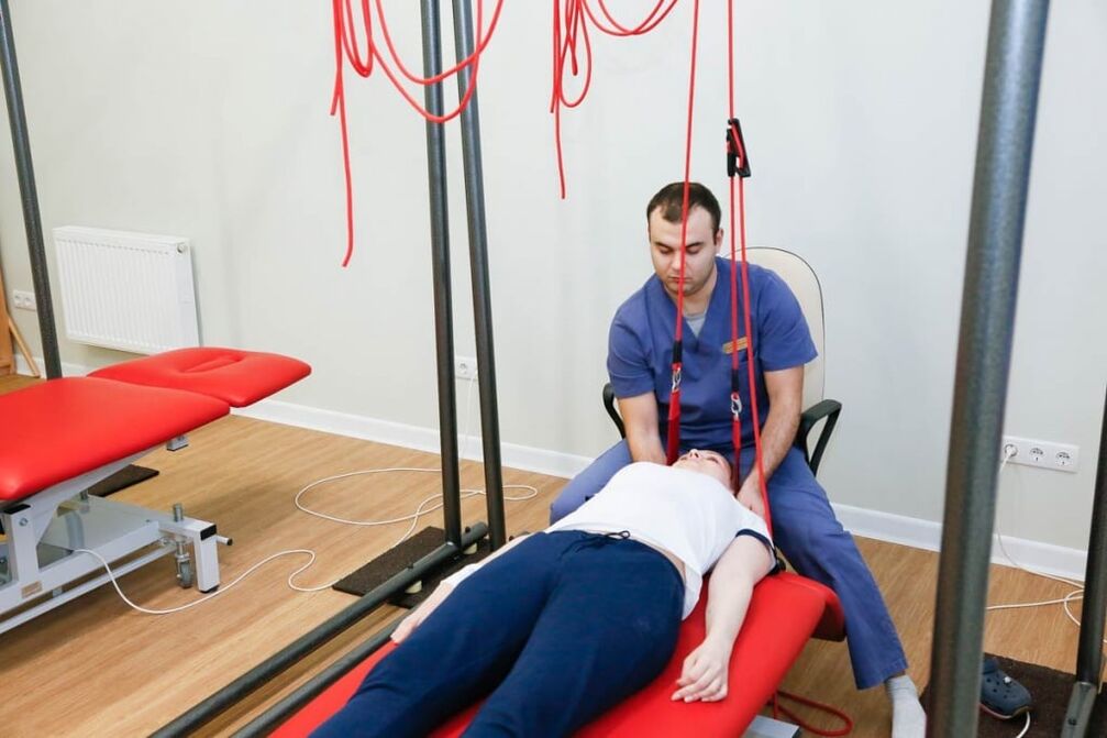
Autogravity therapy is the real name of the spinal traction procedure. It is done using special tools. The goal of therapy is to reduce muscle spasm and restore proper vertebral position. To avoid complications, spinal traction is performed by a doctor.
To increase blood supply in the pathological focus, relieve swelling and relieve pain, the following physiotherapeutic procedures are used:
- Diadynamic current. During this procedure, using special tools, low -frequency currents are applied, which stimulate the muscles, relieving cramps and pain. Has a positive effect, increases tissue trophism;
- Ultraviolet irradiation. Under the influence of UV radiation, vitamin D metabolism improves, calcium content increases, bone tissue becomes stronger;
- Exposure to ultrasound - used to accelerate blood flow, antispasmodic and reparative action. Ultrasound is capable of penetrating deep into tissues, sometimes it is used for better absorption of medicinal substances;
- Amplipulse Therapy - allows you to relieve pain by blocking nerve impulses from the painful focus.
In the acute period of the disease, which lasts 4-7 days, painkillers, antispasmodics, irritants are used to reduce pain. The patient is given peace of mind. Immobilization of the cervical spine was performed using a Shants collar. Exercise therapy and massage are contraindicated. Apply ultraviolet radiation.
The duration of the subacute period was 29 days. Upon full recovery, the patient must rest for a few days. Then you can start a course of rehabilitation therapy. In the chronic process of the disease, patients are given muscle relaxants, chondroprotectors, B vitamins, for pain - analgesics, NSAIDs. Physiotherapy exercises, massages are provided. The patient is discharged physiotherapeutic procedures (amplipulse, alternating current exposure), spinal traction is performed.

Food
Proper nutrition for osteochondrosis is an important condition for achieving remission. The development of cervical osteochondrosis stops with diet and treatment. Neurologists know how to treat osteochondrosis of the cervical spine, therefore, they form a complex of therapeutic measures, including procedures, exercise therapy, proper nutrition and lifestyle changes.
Many patients turn to a neurologist with the question of how to treat cervical spine osteochondrosis and whether there are dietary restrictions. Specialists create individual nutrition programs that take into account patient preferences. The diet for osteochondrosis is based on a balanced and low -fat diet rich in nutrients. The patient's daily diet includes foods high in calcium.
How to sleep with cervical osteochondrosis
For patients with diseases of the musculoskeletal system, the question of how to sleep properly with cervical osteochondrosis is relevant. Sleeping on the stomach provokes the development of the disease, so it is better to avoid sleeping in this position. The most optimal position is on the back and sides.
Cervical osteochondrosis develops while resting in bed with a soft mattress. Therefore, experts recommend giving priority to elastic mattresses, as well as soft pillows. If a patient is diagnosed with cervical osteochondrosis, an experienced specialist will tell you a safe bed to sleep on.
Prophylaxis
To prevent the onset or development of cervical osteochondrosis, doctors recommend:
- Maintain proper body posture;
- Follow an active lifestyle, relax at work;
- Perform regular physiotherapy exercises;
- Sleep on firm and flat surfaces, orthopedic mattresses and pillows;
- Get rid of bad habits, especially smoking;
- Choose shoes taking into account the physiological structure of the foot;
- Do not carry the bag on one hand, this leads to flexion of the spine;
- Live a healthy lifestyle, eat right, eat lots of fruits and vegetables;
- Do not sit long with your head tilted;
- Go swimming.
To improve blood circulation, massage therapy should be done regularly.